【人気ダウンロード!】 e coli under microscope 400x 316490-How does e coli look under a microscope
E coli under the microscope Escherichia coli (E coli) is a bacterium commonly found in various ecosystems like land and water Most of the strains of E coli are harmless, but some strains are known to cause diarrhea and even UTIs E coli is commonly studied as they are considered as a standard for the study of different bacteria(attempting to use the 400X ruler shows this point nicely) 9 But we know that the 400X has ¼ the field of view of the 100X 10 OK, now we can use the higher 400X magnification to make a better estimate of the size of the organism Under the 400X view it takes 3 lengths of the organism to span the diameter of the field of view 11 SOMany bacteria look like E coli when examined under the microscope (if not stained Enterobacteriaceae, Bacillus, cornyeforme
Www Mccc Edu Hilkerd Documents Bio1lab3 Exp 4 Pdf
How does e coli look under a microscope
How does e coli look under a microscope-4 Can a light microscope see bacteria?E Coli E Coli under the microscope at 400x E Coli (Escherichia Coli) is a gramnegative, rodshaped bacterium Most E Coli strains are harmless, but some serotypes can cause food poisoning in their hosts The harmless strains are part of the normal flora of the gut Learn more about E Coli here Helicobacter Pylori


Bacteria Under The Microscope E Coli And S Aureus Youtube
Ecoli is usually motile in liquid or semisolid environment with peritrichous flagella (about 6 per cell) and its surface is covered with fimbriae These structures (flagella and fimbriae) are too thin to be visualized by classical light microscopy or they don't have to be present at all under given cultivation conditions even at motile strainsExamples of Bacteria Under the Microscope Escherichia coli Escherichia coli (Ecoli) is a common gramnegative bacterial species that is often one of the first ones to be observed by students Most strains of Ecoli are harmless to humans, but some are pathogens and are responsible for gastrointestinal infections They are a bacillus shapedMost E Coli strains are harmless, but some serotypes can cause food poisoning in their hosts Depending on the lacto it might be difficult to see at 400x without staining and under bright field As for the lactobacillus fermentum, the shape is rod shapemostly different length and the colour are transluscent
E Coli E Coli under the microscope at 400x E Coli (Escherichia Coli) is a gramnegative, rodshaped bacterium Most E Coli strains are harmless, but some serotypes can cause food poisoning in their hosts The harmless strains are part of the normal flora of the gut Learn more about E Coli here Helicobacter PyloriNucleus, cell membrane,cytoplasm What's missing in ecoli?Cells that have DNA, NO nucleus Exbacteria A rectangular piece of glass where a sample is mounted for viewing under the microscope What is the eye piece?
Yes, most of the bacteria range from 022 µm in diameter The length can range from 110 µm for filamentous or rodshaped bacteria The most wellknown bacteria E coli, their average size is ~15 µm in diameter and 26 µm in length As we talked above, you can see some bacteria in my cheek cellsEscherichia coli or Ecoli is a gramnegative species of bacillus shaped bacteria that can be easily observed under a microscope, even for those with the untrained eye This bacteria has a fast growth rate, doubling every minutes, making them a common choice for bacterial related research purposesE coli under microscope 400x › e coli e coli under microscope oil immersion E Coli Under Microscope Written By MacPride Friday, June 14, 19 Add Comment Edit British E Coli Cases Rise World News The Guardian Microscopic Studies Of Various Organisms


What Does An E Coli Bacteria Look Like Under A Microscope Quora


Gram Stain Demonstration Slide 400x 1 Marc Perkins Photography
Both of bacteria, EColi and S Pyogenes, were observed under the compound light microscope, which is a type of microscope using visible light and a system of lenses to magnify the images In this second lab experiment, four slides of bacteria were viewed under the compound light microscope from 40X up to 400X total magnification for the purpose of giving the images of bacteria in detail beautifullyEscherichia coli bacteria on blood agar e coli under a microscope stock pictures, royaltyfree photos & images petri dishes with culture media for sarscov2 diagnostics, test coronavirus covid19, microbiological analysis e coli under a microscope stock pictures, royaltyfree photos & imagesWhich bacteria look similar to E coli under 100X optical microscope?


Staphylococcus Aureus Under Microscope Microscopy Of Gram Positive Cocci Morphology And Microscopic Appearance Of Staphylococcus Aureus S Aureus Gram Stain And Colony Morphology On Agar Clinical Significance


Observing Bacteria Under The Light Microscope Microbehunter Microscopy
4 Observe Click on the cow and observe E coli under the highest magnification Notice the microscope magnification is larger for this organism, and notice the scale bar is smaller A What is the approximate size of E coli?E Coli E Coli under the microscope at 400x E Coli (Escherichia Coli) is a gramnegative, rodshaped bacterium Most E Coli strains are harmless, but some serotypes can cause food poisoning in their hosts The harmless strains are part of the normal flora of the gut Learn more about E Coli here Helicobacter PyloriObserve Select the human skin sample On the MICROSCOPE tab, choose the 400x magnification, focus on the sample, and turn on Show labels Click on the Nucleus label If necessary, adjust the Stage sliders to see the full description


Lab Manual Exercise 1


Microscope World Blog Staphylococcus Under The Microscope
Fine Focus Which structures are in human skin cells?Get a second escherichia coli bacteria (e coli) stock footage at 2997fps 4K and HD video ready for any NLE immediately Choose from a wide range of similar scenes Video clip id Download footage now!Which bacteria look similar to E coli under 100X optical microscope?


Gram Stain


Bacteria Under The Microscope E Coli And S Aureus Youtube
Which focus is easier for 400x?1 Before you plug in the microscope, turn the light intensity control dial on the righthand side of the microscope to 1 Now plug in the microscope and turn it on (see Fig 6) 2 Place a rounded drop of immersion oil on the area of the slide that is to be observed under the microscope, typically an area that shows some visible stainTo observe E coli with any detail, you will need to use the 100X lens, which is also known as an oil immersion lens This is the longest, most powerful and most expensive lens on the microscope, requiring extra care when using it As the name implies, the 100X lens is immersed in a drop of oil on the slide
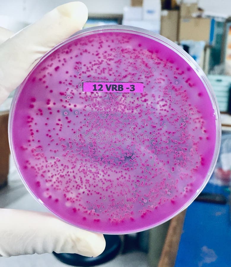


1 735 Microbiology Pink Photos Free Royalty Free Stock Photos From Dreamstime
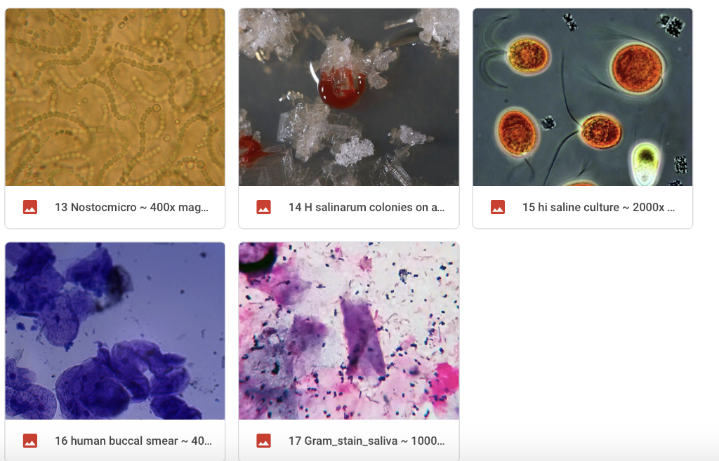


Solved Look At The 2 Gram Stain Slide This Is A Gram Sta Chegg Com
Bacteria E coli Must observe under 400x Very small & motile Looks like specks of sand Hard to discern shape Smaller than yeast & protozoa Instructor to provide demonstration & instructionsMicroscope World recently took a Gram stain prepared slide of three different types of bacteria and captured images using different microscope objective lenses The microscope used to view the bacteria was the Fein Optic R0 biological lab microscopeGram Bacillus Gram positive rod for comparison = yeast (unstained) at 400X total mag


E Coli Video Youtube
.jpg)


Escherichia Coli 400x Escherichia Coli 400x Manufacturers Escherichia Coli 400x Suppliers Escherichia Coli 400x Exporters Escherichia Coli 400x In India
The total magnification of the microscope is calculated by multiplying the magnification of the objectives, with the magnification of the eyepiece Most educationalquality microscopes have a 10x (10power magnification) eyepiece and three objectives of 4x, 10x & 40x to provide magnification levels of 40x, 100x and 400xEscherichia coli Gram stained smear under microscopeRod shapedpink in colorthat's why Gram negative Bacilli#GramStain#GNB#GNREscherichia coli under the microscope if you forgot to apply safranin colorless Escherichia coli under the microscope if you forgot to apply decolorizer purple Bacillus megaterium under the microscope if you forgot to apply iodine pink THIS SET IS OFTEN IN FOLDERS WITH AcidFast Staining 9 terms mafalda5
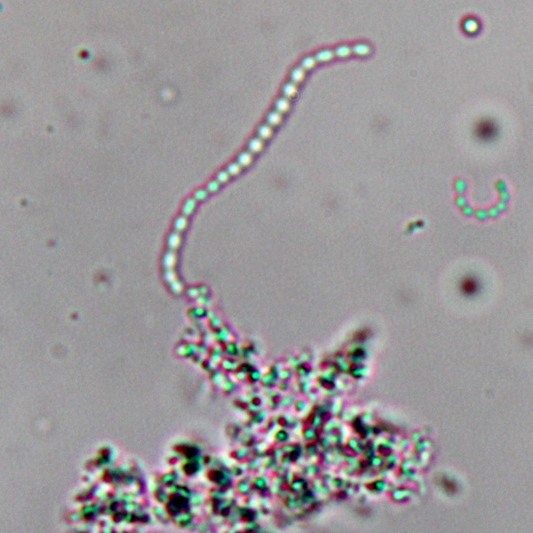


Observing Bacteria Under The Light Microscope Microbehunter Microscopy


Q Tbn And9gcqkye60ou Johpr02n Mbv1fferrjpdh Lnct7ymdf5qhyia1ld Usqp Cau
Nucleus What are prokaryotic cells?Escherichia coli light microscopy Escherichia coli Magnification 1000×E Coli E Coli under the microscope at 400x E Coli (Escherichia Coli) is a gramnegative, rodshaped bacterium Most E Coli strains are harmless, but some serotypes can cause food poisoning in their hosts The harmless strains are part of the normal flora of the gut Learn more about E Coli here Helicobacter Pylori


Microscopy Gram Staining Microscope World Blog


Science Source Stock Photos Video Escherichia Coli And Staph Aureus
E Coli E Coli under the microscope at 400x E Coli (Escherichia Coli) is a gramnegative, rodshaped bacterium Most E Coli strains are harmless, but some serotypes can cause food poisoning in their hosts The harmless strains are part of the normal flora of the gut Learn more about E Coli here Helicobacter PyloriMany bacteria look like E coli when examined under the microscope (if not stained Enterobacteriaceae, Bacillus, cornyeformeCell wall, cell membrane, cytoplasm, flagellum, pilus, and nucleoid C What organelle is missing from E coli?


Royalty Free Escherichia Coli Bacteria E Coli Under Stock Video Imageric Com
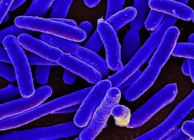


E Coli Under The Microscope Types Techniques Gram Stain Hanging Drop Method
Probably 400X, Anabaena "laxa" From the same sample as above, a few weeks later If your microscope As a rule of thumb, the more transparent the changed optical) and electronic, depending on the principle or method of magnification process again Most E Coli strains are harmless, but some serotypes can cause food poisoning in their hostsTo observe the binding ability of E coli clearly under fluorescent microscope (400X magnification), we transformed GBP in green fluorescent E coli In the result, there were green fluorescent E coli (with GBP) binding on the gold chips (Fig B2) compared with the control group there is no E coli binding on the gold chips (Fig B1)E Coli Under Microscope 400x Gram Stain For Films Sigma Aldrich Pin On Pursuit E Coli Bacteria Gram Negative Bacilli Gram Stain Lm X400 Staphylococcus Aureus And Ecoli Under Microscope Microscopy Of Gram Stain Microbiology Images Photographs B Subtilis Gram Stain 29 March 13 Ibg 102 Lab Reports


Escherichia Coli Bacteria E Coli Stock Footage Video 100 Royalty Free Shutterstock


Photomicroscopy Photomicrography For Bacteriological And
Lactobacillus under the microscope at 400x magnification Place the 100X view on your overhead and focus on your screen the objectives on their microscope by determining the field width for Raphidiopsis curvata Mixed with some straight chains that are either more Raphidiopsis or Cylndrospermopsis raciborskii without akinetes or heterocystsGram Bacillus Gram positive rod for comparison = yeast (unstained) at 400X total magE coli under the microscope Escherichia coli (E coli) is a bacterium commonly found in various ecosystems like land and water Most of the strains of E coli are harmless, but some strains are known to cause diarrhea and even UTIs E coli is commonly studied as they are considered as a standard for the study of different bacteria
-lab-2063642447.jpg)


Escherichia Coli 400x Manufacturers Escherichia Coli 400x Exporters And School Educational Lab Suppliers In India
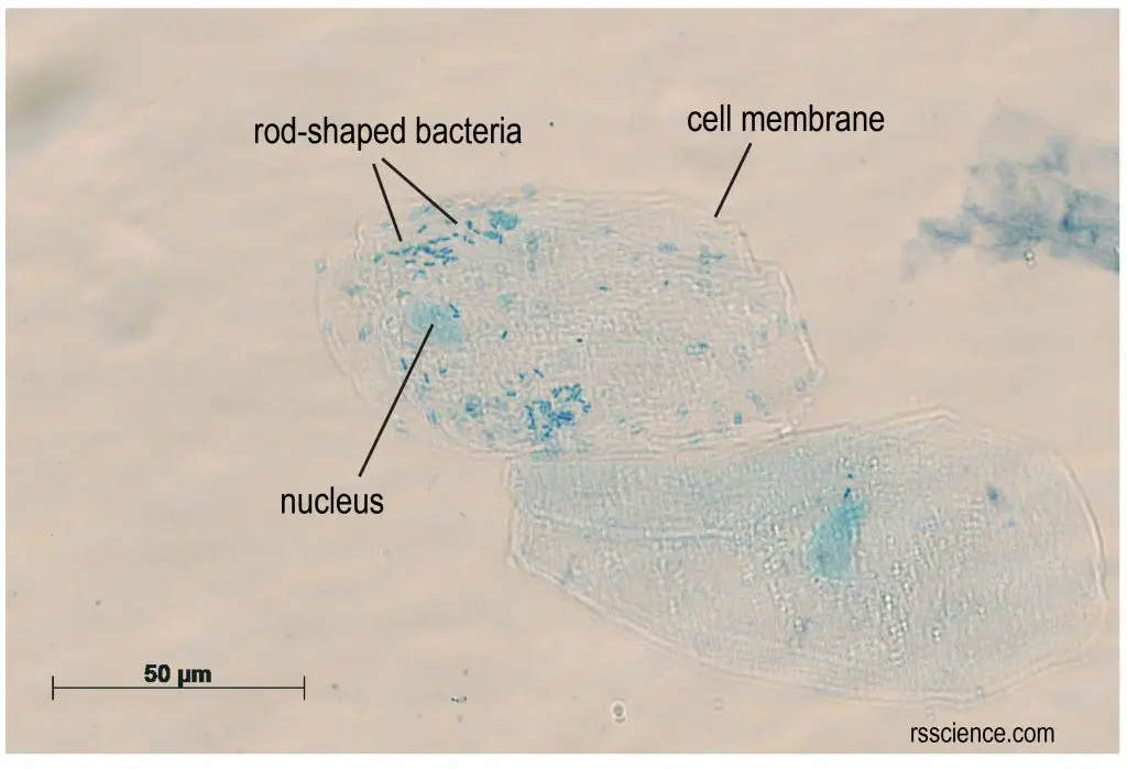


What Living Things You Can See Under A Light Microscope Rs Science
Escherichia Coli (400x) Escherichia coli (E Coli) is one of many species of bacteria that lives in the lower intestines of mammals In the large intestine of mammals, E coli assists in waste processing, vitamin K production and food absorption E coli causes illness in humans Learn more about E coli here Chromosomes (400x)Cheek Cells Under a Microscope Requirements, Preparation and Staining Cheek cells are eukaryotic cells (cells that contain a nucleus and other organelles within enclosed in a membrane) that are easily shed from the mouth lining It's therefore easy to obtain them for observationAbout 15 μm B What organelles are present in E coli?


Observing Bacteria Under The Light Microscope Microbehunter Microscopy
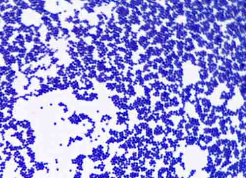


Bacterial Staining Microbiology Images Photographs And Videos Of Gram Acid Fast Endospore
Most E Coli strains are harmless, but some serotypes can cause food poisoning in their hosts Depending on the lacto it might be difficult to see at 400x without staining and under bright field As for the lactobacillus fermentum, the shape is rod shapemostly different length and the colour are transluscentEcoli is usually motile in liquid or semisolid environment with peritrichous flagella (about 6 per cell) and its surface is covered with fimbriae These structures (flagella and fimbriae) are too thin to be visualized by classical light microscopy or they don't have to be present at all under given cultivation conditions even at motile strainsEscherichia coli light microscopy Escherichia coli Magnification 1000×


Www Mccc Edu Hilkerd Documents Bio1lab3 Exp 4 000 Pdf


Www Mccc Edu Hilkerd Documents Bio1lab3 Exp 4 Pdf
E Coli Under Microscope 1000x Written By MacPride Saturday, July 21, 18 Add Comment Edit Chapter 3 Tools Of The Laboratory E Mail Balantidiasis Microscopy Findings Bacteria Bacilli And Cocci Colored With The Gram Staining Flickr Euglena Under Microscope 400x Labeled About;Escherichia coli under the microscope if you forgot to apply safranin colorless Escherichia coli under the microscope if you forgot to apply decolorizer purple Bacillus megaterium under the microscope if you forgot to apply iodine pink THIS SET IS OFTEN IN FOLDERS WITH AcidFast Staining 9 terms mafalda5• A basic light microscope has 4 objective lenses – 4X, 10X, 40X, and 100X The higher the number, the higher the power of magnification Once you have focused the


What Does An E Coli Bacteria Look Like Under A Microscope Quora
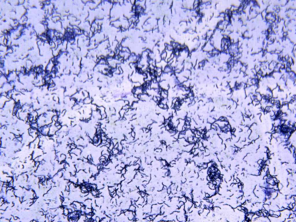


Solved In This Section You Will View A Random Set Of Micr Chegg Com
Escherichia coli under the microscope if you forgot to apply safranin colorless Escherichia coli under the microscope if you forgot to apply decolorizer purple Bacillus megaterium under the microscope if you forgot to apply iodine pink THIS SET IS OFTEN IN FOLDERS WITH AcidFast Staining 9 terms mafalda5Microbes under the microscope STUDY PLAY Escherichia Coli Bacillus anthracis Trichomonas vaginalis Escherichia Coli infoBacteriaFacultative anaerobeEndospore formingGram positiveRodshaped 400XChemoeterotrophSpore FormingFermentationOpportunistic pathogenSource of PenicillinWhat Does An E Coli Bacteria Look Like Under A Microscope Quora If you have a microscope 400x and a properly stained slide of the onion root tip or allium root tip you can see the phases in different cel Diagram Compound Light Microscope Parts Labeled


Escherichia Coli Bacteria E Coli Stock Footage Video 100 Royalty Free Shutterstock


Microscopy And Staining
E Coli Under Microscope 400x E Coli Under The Microscope Types Techniques Gram Stain Chapter 3 Tools Of The Laboratory E Mail Bacteria Under The Light Microscope A B Subtilis Hh2 Was E Coli Under Electron Microscope Science Source E Coli Bacteria Light MicroscopyE Coli Under Microscope 400x Gram Stain For Films Sigma Aldrich Pin On Pursuit E Coli Bacteria Gram Negative Bacilli Gram Stain Lm X400 Staphylococcus Aureus And Ecoli Under Microscope Microscopy Of Gram Stain Microbiology Images Photographs B Subtilis Gram Stain 29 March 13 Ibg 102 Lab ReportsWhen viewed under the microscope, Gramnegative E Coli will appear pink in color The absence of this (of purple color) is indicative of Grampositive bacteria and the absence of Gramnegative E Coli Escherichia coli under 10х90х magnification using fuchsine as a dye by ElNokko (Own work) CC BYSA 40 (http//creativecommonsorg/licenses/bysa/40), via Wikimedia Commons


Igem Research Article E Cotector The Fluorescent E Coli With Surface Displayed Anti Cancer Marker Scfv To Detect Specific Cancer Markers Plos Collections


Gram Stain
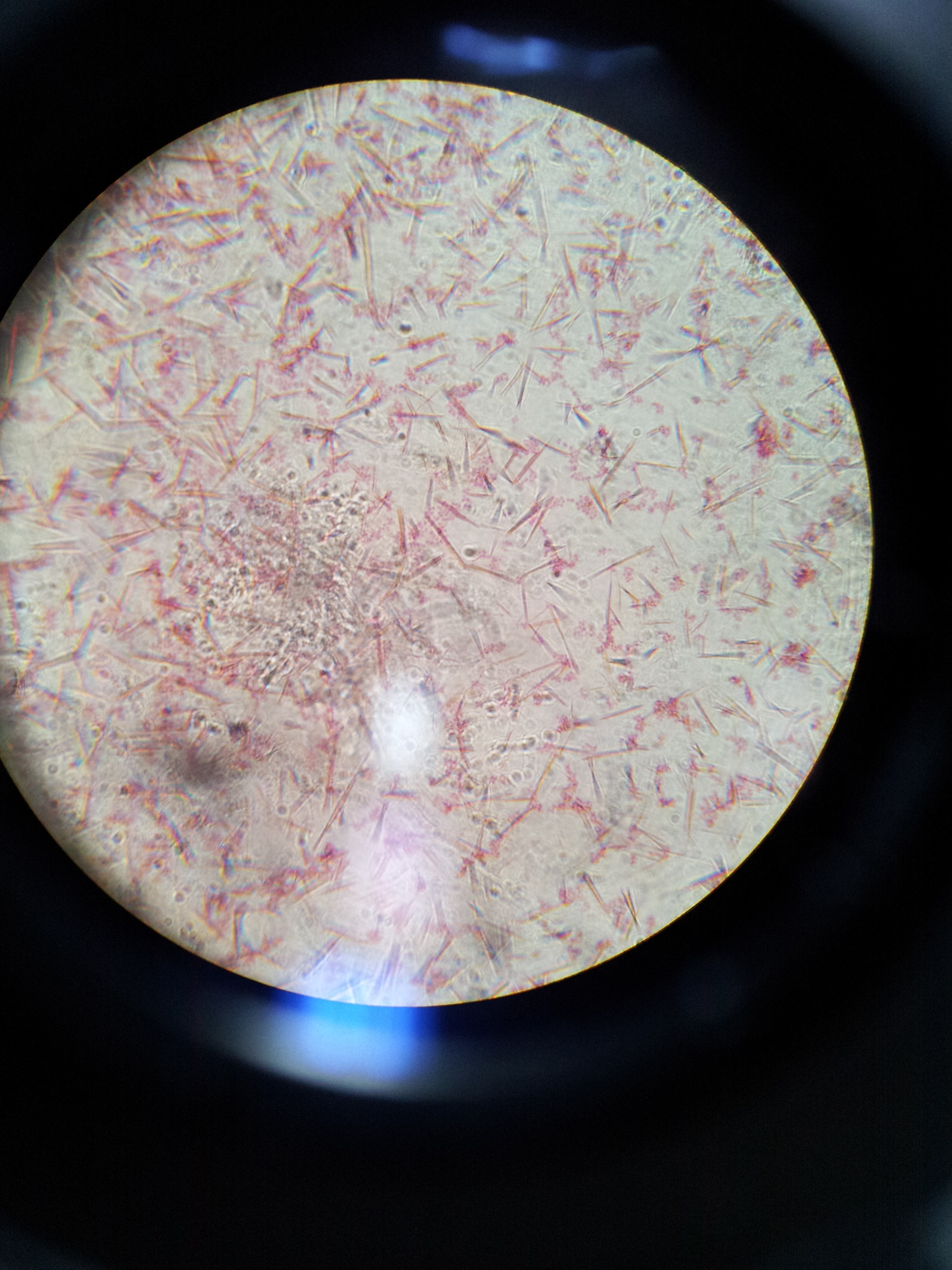


Lab 1 Principles And Use Of Microscope Ibg 102 Lab Reports


1 735 Microbiology Pink Photos Free Royalty Free Stock Photos From Dreamstime


Microscope World Blog Microscopy Gram Staining


Light Microscopy Streaked Images


Gut Bacteria Escherichia Coli Under Microscope Youtube


Team Nymu Taipei Project 1c1 14 Igem Org


Gram Stain


Biol 2


Biol 230 Lab Manual Lab 1


Escherichia Coli Bacteria E Coli Stock Footage Video 100 Royalty Free Shutterstock


Afip Wsc 96 97 Conference 1


Q Tbn And9gcqy Atzkf0po8c8x1yoohusnpjlpu6pnvquhzmi5dmxlldlhkom Usqp Cau


E Coli Bacteria Under Microscope Page 1 Line 17qq Com
.jpg)


Biol 2


Gram Stain
.jpg)


Escherichia Coli 400x Escherichia Coli 400x Manufacturers Escherichia Coli 400x Suppliers Escherichia Coli 400x Exporters Escherichia Coli 400x In India


Cocci Bacteria Under Microscope Page 1 Line 17qq Com


Biol 230 Lab Manual Lab 1


49 Under The Microscope Ideas In 21 Microscopic Photography Things Under A Microscope Microscopic
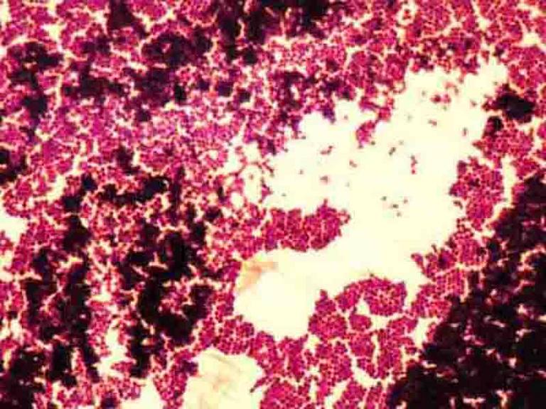


Gram Stain Microbiology Images Photographs
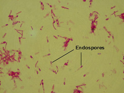


Micromorphology Slides Microbiology Resource Center Truckee Meadows Community College
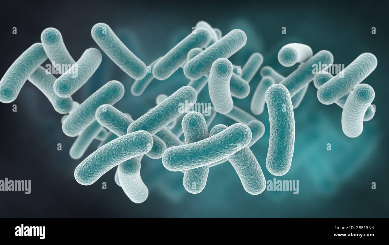


Rod Shaped Bacteria High Resolution Stock Photography And Images Alamy


Structure And Function Of Bacterial Cells


Science Source Stock Photos Video Escherichia Coli And Staph Aureus
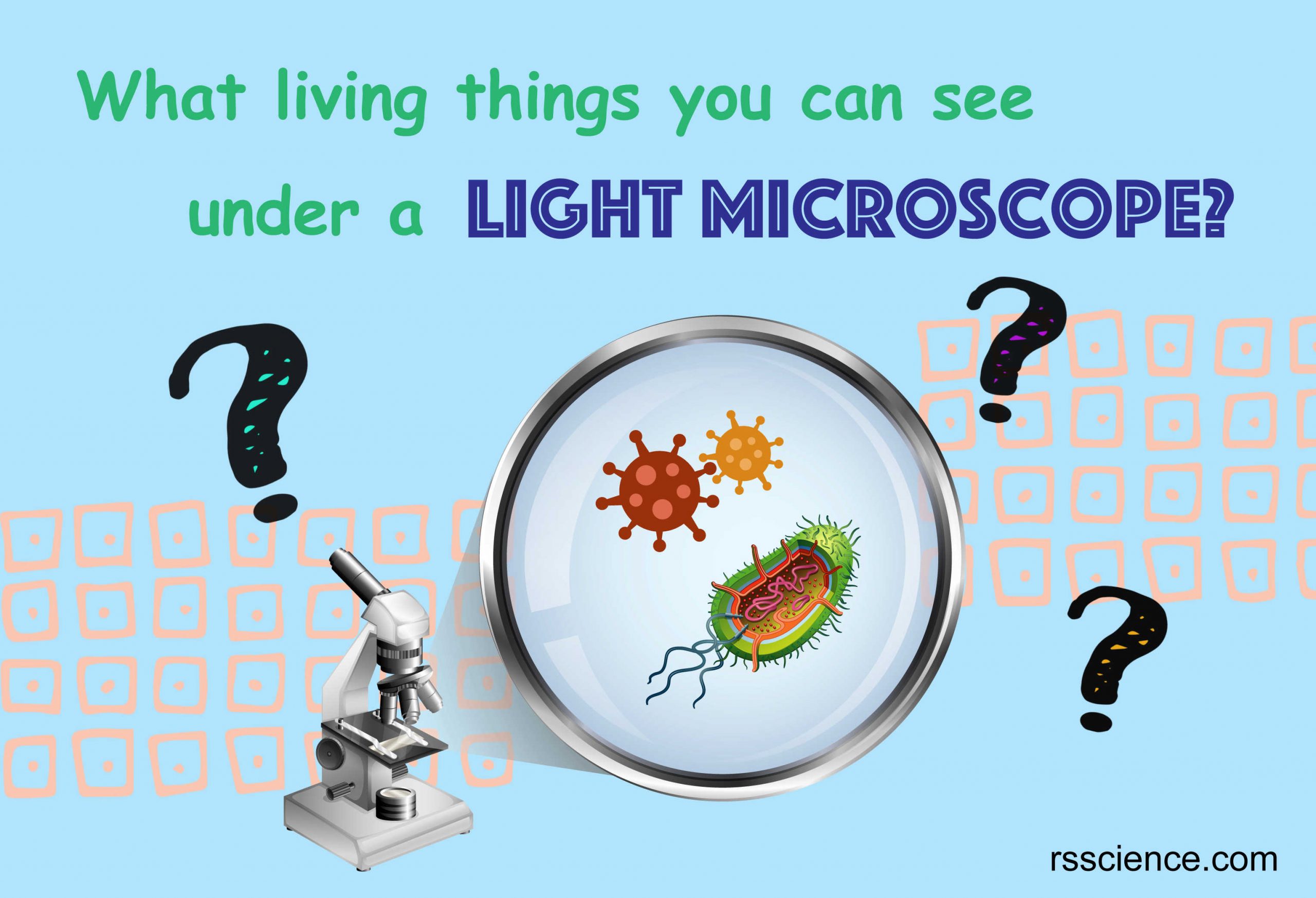


What Living Things You Can See Under A Light Microscope Rs Science


Tissue Sections Light Microscopy H E Stain A C 0x Magnification Download Scientific Diagram
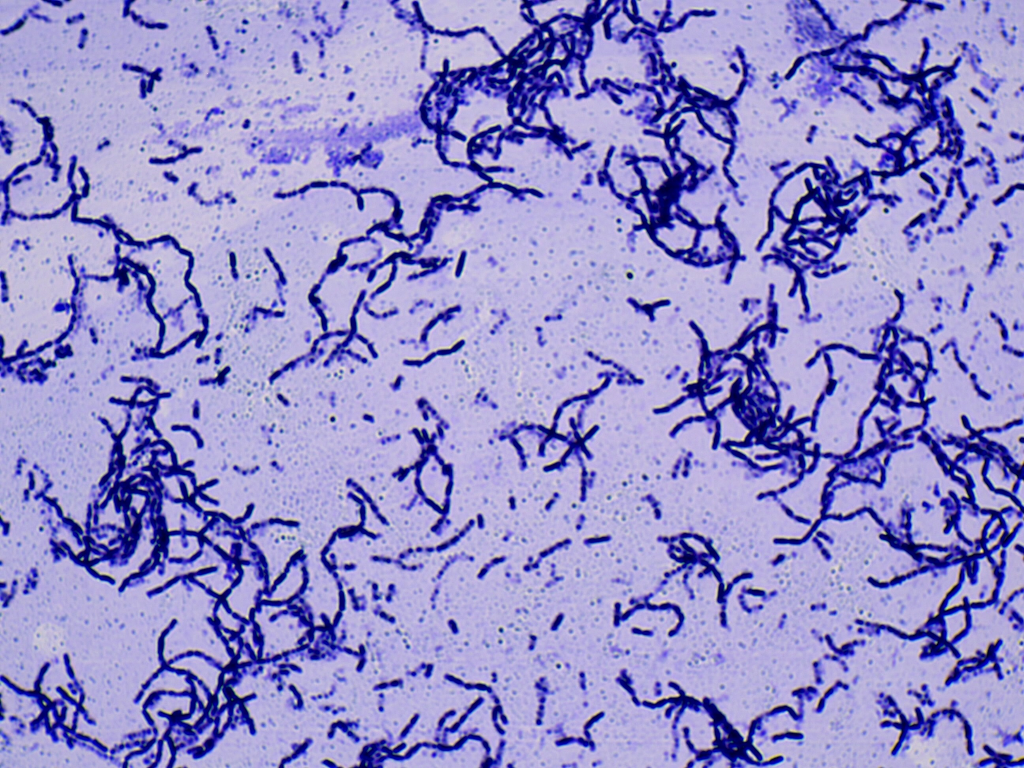


Solved In This Section You Will View A Random Set Of Micr Chegg Com
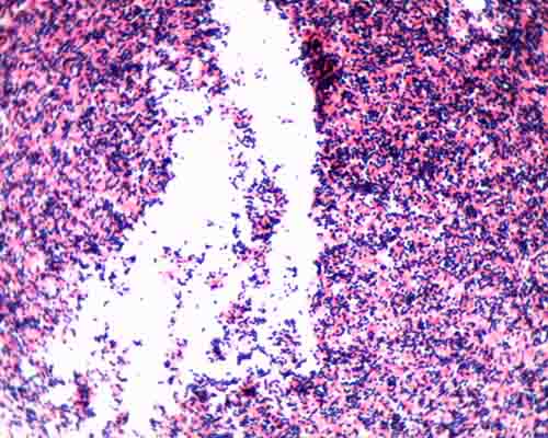


Gram Stain Microbiology Images Photographs


The World S Best Photos Of Prokaryote Flickr Hive Mind Prokaryotes Microbiology Microbiology Study


Biol 230 Lab Manual Lab 1


What Does An E Coli Bacteria Look Like Under A Microscope Quora


What Does An E Coli Bacteria Look Like Under A Microscope Quora


Pin On E Coli
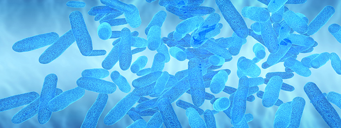


What Magnification Do I Need To See Bacteria Westlab


Team Ustc China Project Results 14 Igem Org


Gram Stain Demonstration Slide 400x 2 A Slide Demonstrati Flickr


Thomasthinktank Licensed For Non Commercial Use Only Observing Microbes Lab


Microscope World E Coli Captured Under A Microscope At 400x Magnification Http T Co T8fnqganhv
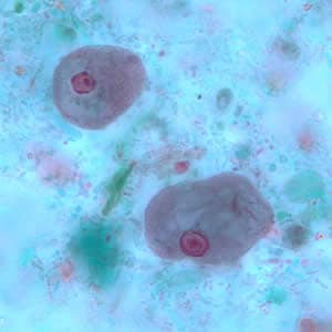


Cdc Dpdx Intestinal Non Pathogenic Amebae


Figure 1 From Detection Of Multidrug Resistant And Shiga Toxin Producing Escherichia Coli Stec From Apparently Healthy Broilers In Jessore Bangladesh Semantic Scholar


E Coli Bacteria Under Microscope Page 1 Line 17qq Com
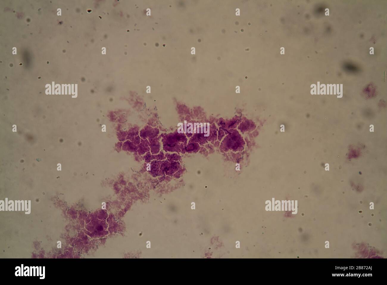


Streptococcus High Resolution Stock Photography And Images Alamy


Observing Bacteria Under The Light Microscope Microbehunter Microscopy


Microscopy And Staining


E Coli Bacteria Under Microscope Page 1 Line 17qq Com
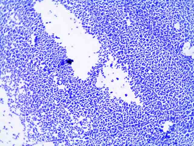


Bacterial Staining Microbiology Images Photographs And Videos Of Gram Acid Fast Endospore


Light Microscopy Streaked Images


Www Mccc Edu Hilkerd Documents Bio1lab3 Exp 4 000 Pdf


Magnification Bioninja
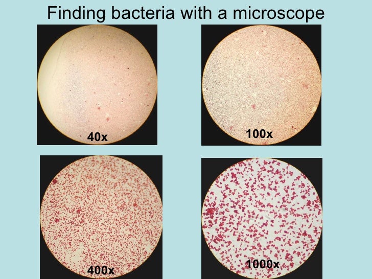


Chapter 3 Tools Of The Laboratory E Mail


Biol 230 Lab Manual Lab 1
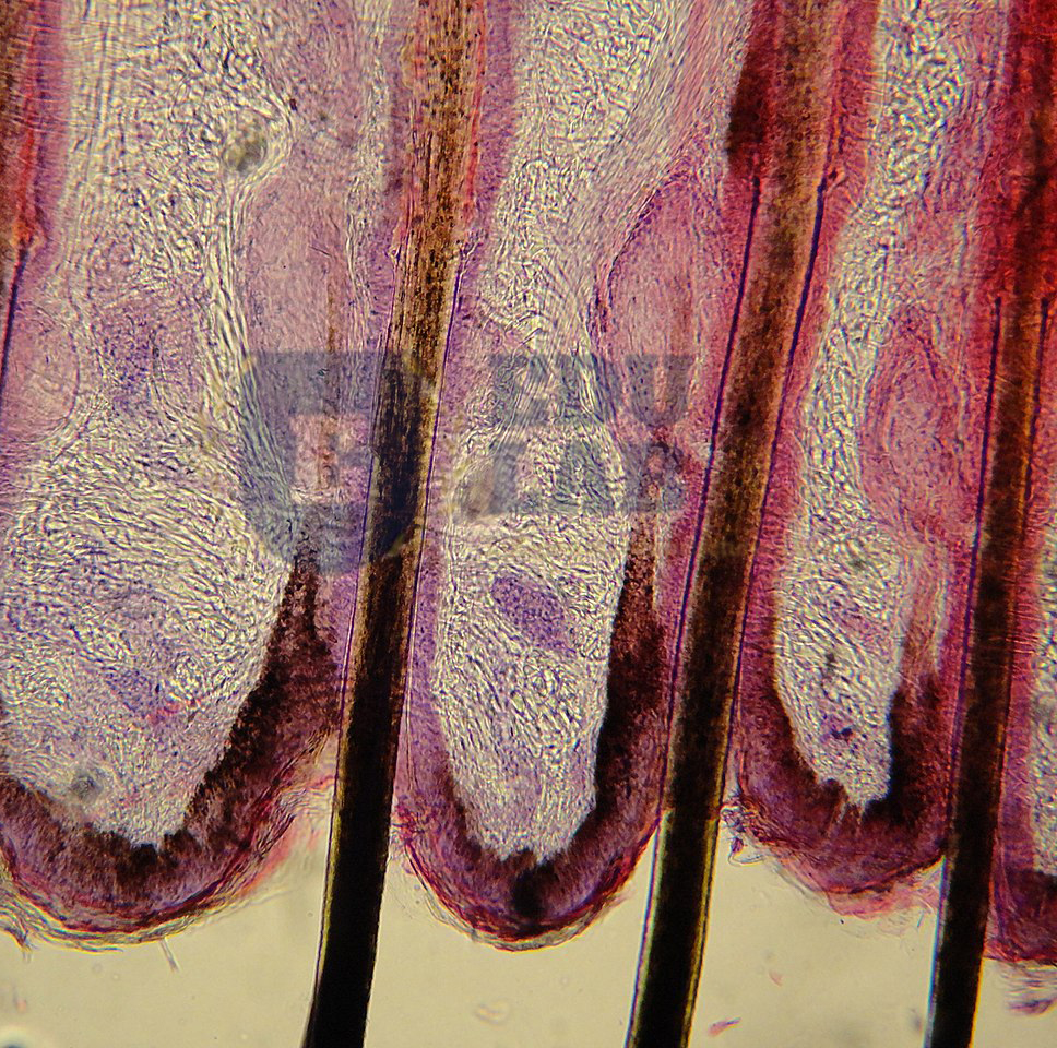


Escherichia Coli 400x Escherichia Coli 400x Manufacturers Escherichia Coli 400x Suppliers Escherichia Coli 400x Exporters Escherichia Coli 400x In India


Secreted Autotransporter Toxin Sat Induces Cell Damage During Enteroaggregative Escherichia Coli Infection
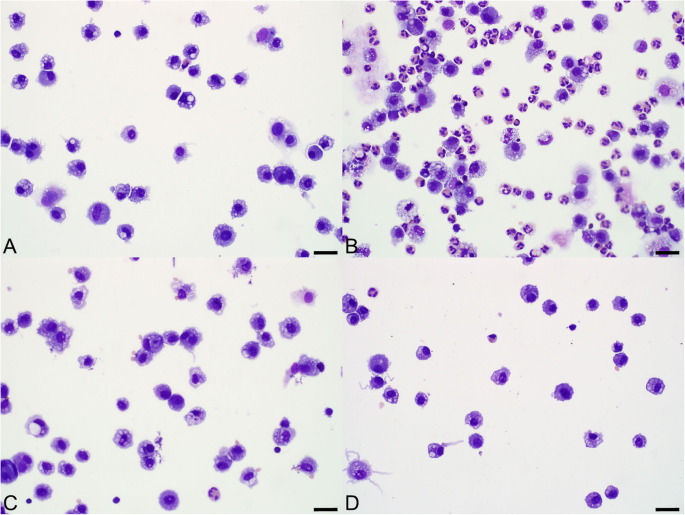


Pulmonary And Systemic Responses To Aerosolized Lysate Of Staphylococcus Aureus And Escherichia Coli In Calves Bmc Veterinary Research Full Text


Q Tbn And9gcs3nlev0tx1pfsddwm6y9ajujk4lxjzmg7ksdxctgnipv0l50c Usqp Cau


E Coli Negative Stain Page 1 Line 17qq Com


What Does An E Coli Bacteria Look Like Under A Microscope Quora


Colour Online Invasive Pattern Of The Escherichia Coli O96 H19 Download Scientific Diagram


Microbes Under The Microscope Flashcards Quizlet


Escherichia Coli Light Microscopy


1 276 Bacillus Subtilis Photos And Premium High Res Pictures Getty Images
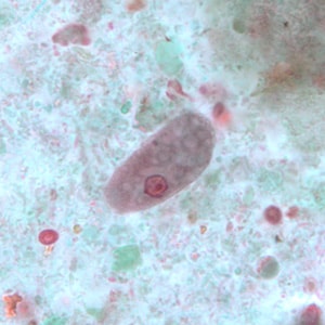


Cdc Dpdx Intestinal Non Pathogenic Amebae
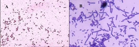


How These 26 Things Look Like Under The Microscope With Diagrams


What Does An E Coli Bacteria Look Like Under A Microscope Quora


Light Microscopy Streaked Images


Aph162 Report 1


Q Tbn And9gctqlwezc G Rsexb5gmw Uv65za98k6p92dhlvblkp4mnrofyo Usqp Cau


Microscope World Blog June 15


347 Gram Stain Photos And Premium High Res Pictures Getty Images
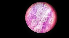


E Coli Under The Microscope Types Techniques Gram Stain Hanging Drop Method
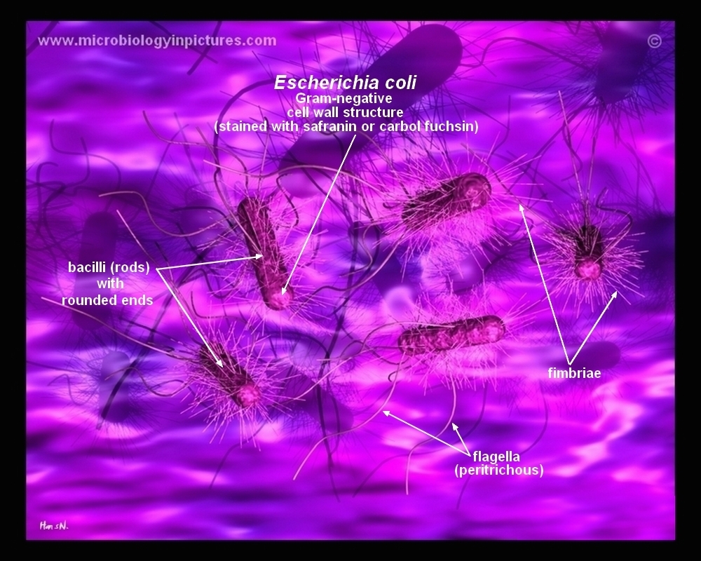


How E Coli Bacteria Look Like


Is A 1 000x Zoom On A Microscope Enough To See Bacteria Cells Quora


コメント
コメントを投稿